| Home | AIDS | What is...? | Gallery | Useful Links | Glossary |
Aids - HIV gallery
|
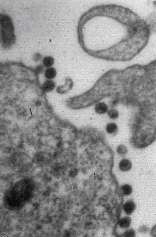
1. Human immunodeficiency virus, viral particles are seen at low magnification adjacent to the cell surface in this electron micrograph.
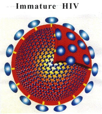
2. Immatured HIV
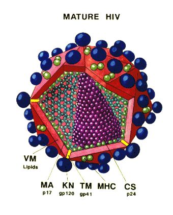
3. Matured HIV
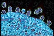
4. Pseudocolored transmission electron micrograph of human immunodeficiency virus (HIV) on infected human lymphocyte. Observe the daughter hiv cells leave the infected t-cell for a new host.
5. from the exHIV(http://www.robin.ru/kahn/) website by Dolph Kahn, Russia
|